Researchers develop artificial blood vessels using technique called co-SWIFT
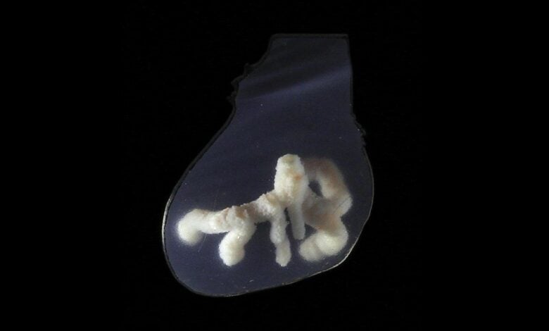
Researchers have made significant strides in developing artificial blood vessels using a technique called co-SWIFT, which involves 3D printing a network of blood vessels within functional human cardiac tissue, Advanced Science News reported.
These artificial vessels feature a dual layer of cells—endothelial and smooth muscle—mimicking the structure of natural blood vessels. When a blood-like liquid was introduced into this network, the cardiac tissue began to contract synchronously, effectively simulating the beating of a heart.
The functionality of these artificial blood vessels was further demonstrated by administering cardiac drugs. For example, isoproterenol, a medication used for treating bradycardia, caused the cardiac tissue to beat twice as fast, while blebbistatin, which inhibits heart cell contraction, halted the tissue’s beating altogether. This ability to respond to drugs indicates that the engineered tissues can mimic real cardiac responses.
Moreover, the researchers successfully printed a replica of a patient’s left coronary artery based on imaging results, showcasing co-SWIFT’s potential for creating patient-specific vascular solutions. This capability is crucial for advancing personalized medicine and highlights the technique’s relevance in generating biologically relevant blood vessels within functional human tissues.